In the field of healthcare, precision, collaboration, and strategy are vital. Without a clear communication process, structured checkpoints, and a well-defined approach, even the most advanced medical initiatives can face setbacks. The Hare Krishna Foundation Trust stands as a beacon of excellence, ensuring that every aspect of medical imaging and diagnostics is handled with utmost accuracy and care.
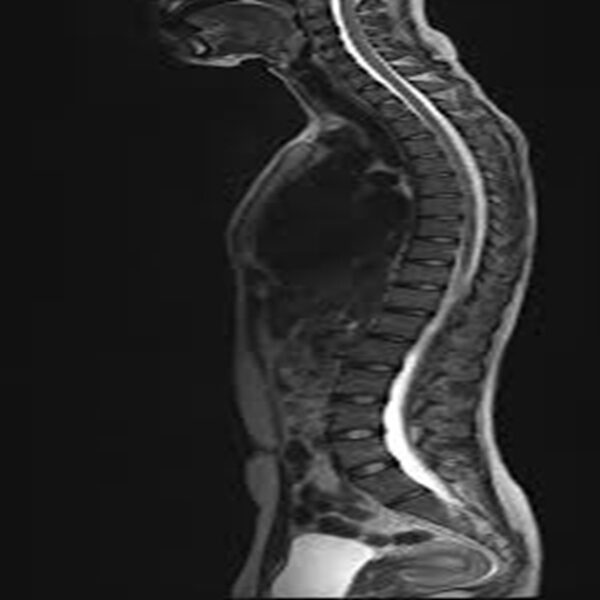
The foundation operates with a mission to provide high-quality imaging services that aid in accurate medical diagnoses and treatments. Our state-of-the-art imaging center is equipped with advanced diagnostic tools, including MRI, CT scan, ultrasound, and digital X-ray technologies, ensuring that patients receive precise and timely results.
Excellence in Medical Imaging
At Hare Krishna Foundation Trust, we believe that a strong diagnostic foundation is essential for effective treatment. Our imaging center follows strict quality control measures, ensuring accuracy in every scan and report. Whether it is a routine health check-up or a critical diagnostic requirement, our services are designed to meet the needs of patients and healthcare professionals alike.
Cutting-Edge Technology & Expertise
We have invested in advanced medical imaging technology to provide high-resolution images that assist doctors in making informed decisions. Our team of experienced radiologists and technicians work diligently to maintain the highest standards of diagnostic imaging.
Why Choose Hare Krishna Foundation Trust?
State-of-the-Art Imaging Equipment: Our center is equipped with the latest medical imaging technology for accurate diagnosis.
Experienced Radiologists: Our team consists of highly qualified radiologists and technicians with years of expertise.
Patient-Centered Approach: We prioritize patient comfort and ensure a smooth, hassle-free experience.
Affordable & Accessible Services: High-quality imaging services at reasonable costs, ensuring healthcare is accessible to all.
Comprehensive Diagnostic Solutions: From routine X-rays to advanced MRI and CT scans, we offer a full spectrum of imaging services.
A Commitment to Quality Healthcare
The Hare Krishna Foundation Trust envisions a future where medical diagnostics are accessible, affordable, and accurate. Our imaging center is more than just a facility—it is a commitment to excellence, ensuring that every diagnosis is backed by reliable technology and expert analysis.
For appointments and inquiries, visit our imaging center or contact us today. Let us be your trusted partner in medical diagnostics and healthcare excellence.


Reviews
Clear filtersThere are no reviews yet.