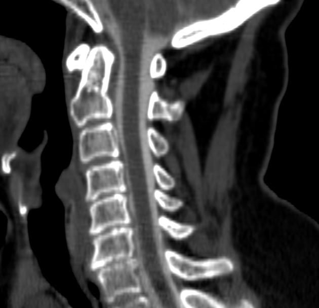
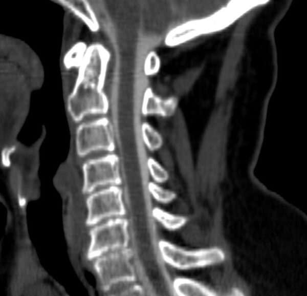
Myelography is a specialized X-ray or CT imaging procedure used to examine the spinal cord, nerve roots, and spinal canal, particularly when evaluating conditions that affect the spinal cord or surrounding structures. It is especially useful in detecting spinal cord compression, herniated discs, tumors, infections, arachnoiditis, or nerve root abnormalities. In this procedure, a contrast dye (iodine-based) is injected into the subarachnoid space—the area around the spinal cord that contains cerebrospinal fluid—through a lumbar puncture (spinal tap), usually in the lower back. After the injection, X-rays and/or CT scans are taken to track the flow of the contrast medium and visualize the outline of the spinal cord and nerve roots. The dye helps highlight abnormal pressure points, blockages, or structural deformities. The patient is often tilted in different directions on the imaging table to allow the contrast to spread evenly throughout the spinal canal. The test is usually performed when MRI is not available or not suitable, especially for patients with metal implants or pacemakers. While the procedure may cause temporary headache or discomfort, it is generally safe when performed with proper precautions. After the exam, the patient is monitored and advised to rest with their head elevated, stay hydrated, and report any unusual symptoms. Myelography provides highly detailed images of the spinal anatomy, helping doctors accurately diagnose the cause of back pain, limb numbness, or weakness, and is often used in pre-surgical planning or when MRI results are inconclusive.
Menu


Reviews
Clear filtersThere are no reviews yet.